

Understanding the complexities of your cornea is essential for your eye doctor’s understanding of your eye health. Corneal tomography and corneal topography are two powerful diagnostic tools, and while they might sound similar, these technologies offer distinct insights into your vision health.
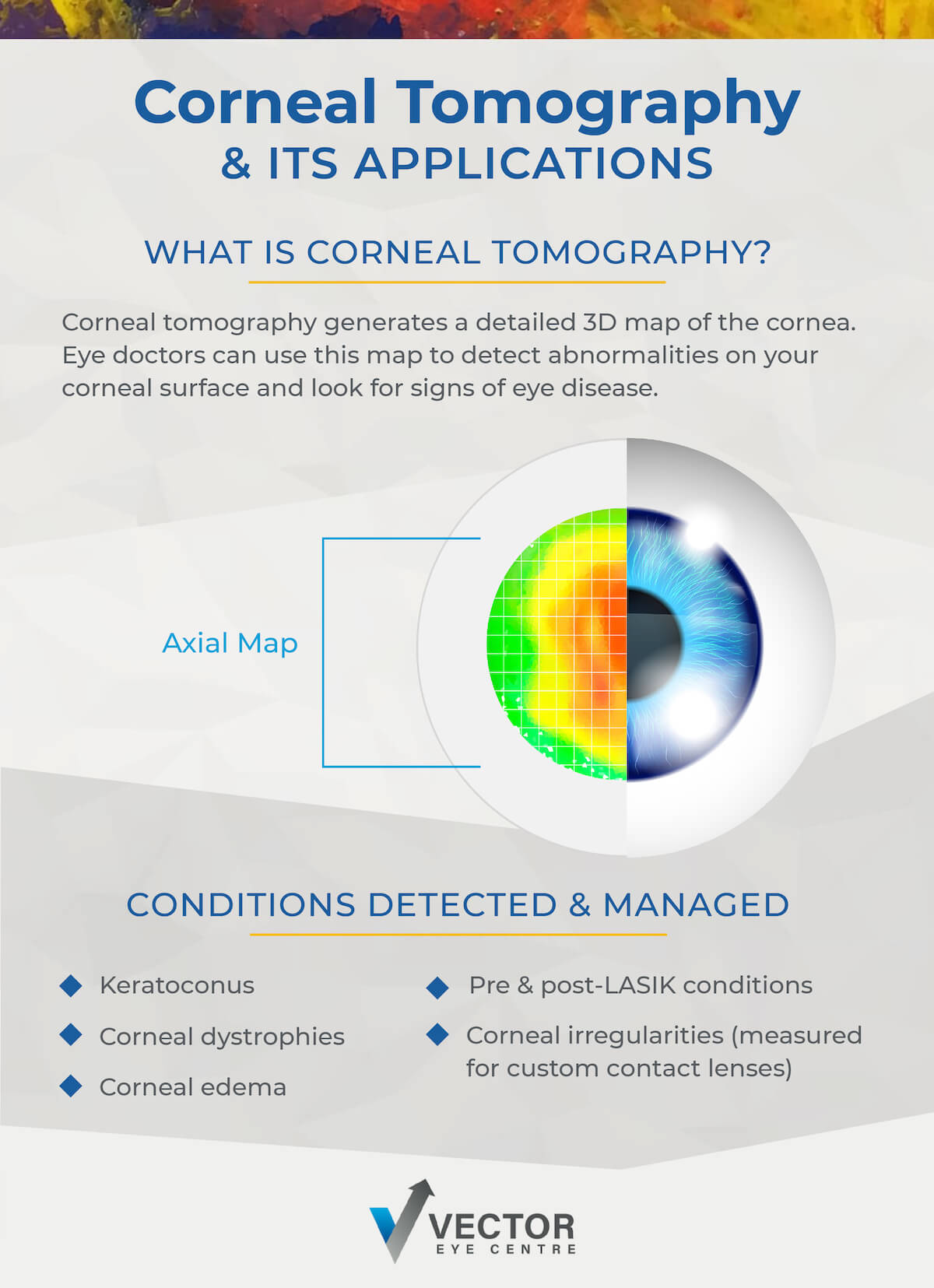
Corneal topography provides a detailed map of the cornea’s surface curvature, highlighting irregularities that may affect vision. Corneal tomography goes deeper by capturing a 3-dimensional image of the cornea, including its thickness, volume, and structural integrity, offering a comprehensive view of corneal health.
Corneal Health & Its Impact on Vision
Your cornea is the eye’s outermost layer, acting as a window controlling light entry. Any irregularities or diseases affecting the cornea can lead to significant vision issues, from mild blurriness to severe, vision-threatening conditions.
Maintaining corneal health is crucial for:
- Clear Vision: A healthy cornea allows light to focus on the retina properly.
- Eye Protection: The cornea acts as a barrier against dirt, germs, and other particles.
- Optimal Eye Function: Proper corneal structure supports overall eye function and health.
- Ocular Comfort: Much of eye discomfort is caused by irregularities of the cornea surface.
Understanding How Corneal Tomography Works
Imagine your eye is like a camera; the cornea is the clear, front lens. The lens must be in good shape for your “camera” to take clear pictures (allow you to see clearly). Corneal tomography is similar to getting a super-detailed, 3D X-ray of that lens but utilizes a very safe method of 3D rotational photography.
This allows your eye doctor to get a complete, inside and out view of your cornea, helping ensure your eyes stay healthy and your vision clear. Here’s a step-by-step look at how it works:
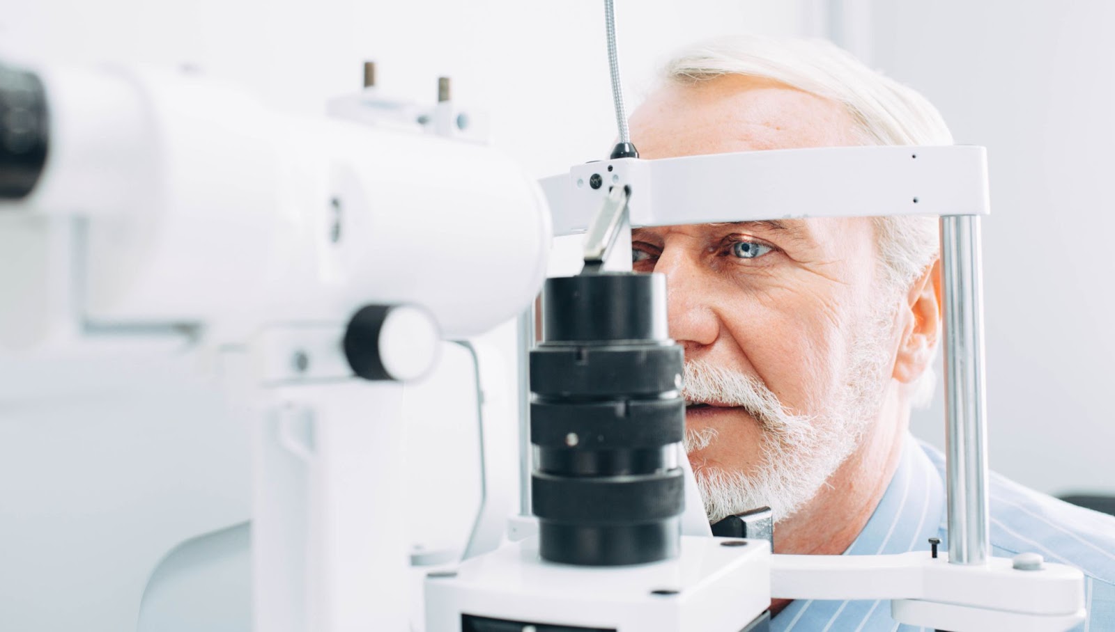
- Preparation: You are seated in front of the tomography device, and your head is positioned correctly.
- Imaging: The device emits a series of light beams that scan your cornea from different angles.
- Data Collection: These light beams capture, through Scheimpflug technology, detailed information about your cornea’s shape, thickness, and internal structures.
- 3D Model Creation: The collected data is processed for a comprehensive 3-dimensional cornea image.
Corneal tomography can provide detailed imaging that can help detect subtle abnormalities that are not visible with traditional examination methods.
Applications of Corneal Tomography in Eye Care
Corneal tomography has many applications in modern eye care, making it an invaluable tool for your eye doctor. Some of its key uses include:
- Preoperative Assessment: Before LASIK or other refractive surgeries, corneal tomography helps assess corneal thickness and stability, supporting your safety and optimal outcomes.
- Keratoconus Detection: Early detection of keratoconus, a condition where the cornea thins and bulges, is critical. Tomography can identify early signs, allowing for timely intervention. We also can use tomography to diagnose pellucid marginal degeneration and keratoglobus.
- Cornea Ectasia: In rare cases, after laser vision correction, the cornea may become destabilized years after surgery, resulting in shape changes and thinning. Tomography can diagnose these changes at early stages to help with preventing progression with CXL.
- Monitoring Corneal Dystrophies & Degenerations: Regular tomography scans can track the progression of corneal dystrophies and other degenerative conditions.
- Contact Lens Fitting: Detailed corneal measurements aid in designing custom contact lenses for those with irregular corneas.
Corneal Tomography for Managing Corneal Conditions
Corneal conditions can range from minor irritations to serious diseases. Corneal tomography provides critical insights that assist in diagnosing and managing these conditions effectively.
Early Diagnosis
With its detailed imaging capabilities, tomography can detect conditions like keratoconus, corneal ectasia, and corneal edema long before they manifest noticeable symptoms.
Timely detection and intervention help slow down or even halt the progression of debilitating corneal diseases. With the high-resolution images and comprehensive data provided by corneal tomography, eye care professionals can make more informed decisions and initiate appropriate treatments early.
Treatment Planning
Tomography offers a clear picture of your cornea’s structure, helping plan surgical or therapeutic treatments tailored to your specific needs.
In cases where surgical intervention is not required, corneal tomography aids in devising therapeutic strategies tailored to your unique corneal structure. For example, in managing keratoconus, tomography helps select the appropriate intervention, such as corneal cross-linking, to stabilize the cornea effectively.
Monitoring Progress
For chronic conditions, regular tomography scans allow for monitoring the effectiveness of treatments and making necessary adjustments over time.
Whether managing chronic conditions like corneal dystrophies or responding to changes in corneal health due to trauma or infection, the detailed insights provided by tomography help treatment plans remain responsive and effective.
Enhancing Patient Education & Engagement
Corneal tomography enhances your education and engagement by providing visual aids that clearly explain your conditions and the rationale behind your treatment plans.
Seeing a 3D model of your cornea helps you understand the necessity of specific treatments and encourages adherence to prescribed therapies or follow-up appointments.
Caring for Your Corneal Health
Corneal tomography represents a significant advancement in eye care, providing comprehensive insights that surpass traditional diagnostic methods. A detailed view of your cornea’s structure aids in early diagnosis, effective treatment planning, and continuous monitoring of various corneal conditions.
At Vector Eye Centre, we are committed to leveraging the latest medical technology to provide you with a high standard of care. We have advanced technologies of tomography and topography and offer the best in cornea diagnosis, treatment and monitoring. If you have concerns about your corneal health or are interested in learning more about corneal tomography, we can help. To become a patient at Vector Eye Centre, contact us to learn more about how we can help.